Skin Cancer Check with Skin Photos
This free skin cancer check online app can identify potential skin cancer from skin photo images or directly from camera of mobile devices. It measures probability of risk or being cancerous for skin lesions and moles. This is software service provided by Artificial Intelligence (AI) Deep Learning Neural Network Models powered by CMSR Machine Learning Studio. This system has very high accuracy. But is NOT 100% accurate! (See Limitations & System Accuracy.) Note that this is not a medical diagnosis system nor clinical screening device. It's a measuring device for skin risk, especially for skin cancer risk. For much accurate medical diagnosis, you may need biopsy and pathological tests. There are limitations what photos can tell you! Visual photo analysis cannot replace biopsy and pathological tests. Certain skin cancer lesions look alike pimple or rash, especially at early stage, and skin infection. This system can make errors. Note that pimple or rash normally improves or disappears in days. Unhealing skin infections which look similar to skin cancer may still require treatment. Therefore if conditions don't improve, please see a doctor or dermatologist for medical diagnosis and treatments. For non mobile touch version, please use Mouse-enabled Computers (Windows, Mac, Linux, RPi).
This online app is not for general public use. It should be used only for testing and research purposes. If you are interested in licensing this technology, please Contact Us. Models are available in web, Java, C/C++/Objective-C and Swift. User Agreements Skin Lesion Conditions |
||||||||
|
|
||||||||
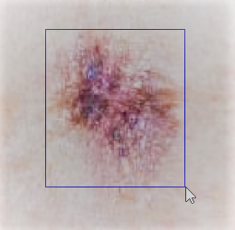
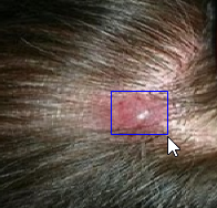
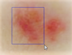
Instructions Take "well-focused" color skin photos. Most Android and iOS web browsers allow you to take photos directly from this software. Skin spots should not be tiny. 5% to 50% of image size is recommended. (For more, please read Photo Requirements.) Tick the skin lesion conditions correctly for each skin lesion. Press the choose file button and select your skin image file. Adjust the rectangular selection box to around a suspected spot or mole. Note that the selection box should be tight but a little wider to the suspected spot or mole as shown in the following figures. Otherwise result can be random! Select the "top and left" option. Swipe the canvas to align the top and left borders. Choose the "bottom and right" option. Swipe the canvas to align the bottom and right borders. If skin lesion is too small, it's difficult to draw a "tight but a little wider" box. Select the "shift image" options to enlarge and move lesions with pinch and swipe operations. Enlarge the skin lesion to occupy at least 10% of the canvas and then adjust the selection box. If skin lesion is not clear, use the "resize canvas" option to adjust the canvas size. (For more, please read Skin Lesion Selection.)


Finally, press the "CHECK" button (or touch the small canvas below the main canvas). Result will be shown below the canvas. Try multiple times with a little larger and smaller selection box areas. Adjust the skin lesion conditions for each lesion if different. When multiple spots are closely together (as in the 3rd above image), try each spot individually as well as together. Results are shown in percentages which indicate skin cancer probabilities, ranging 0% to 100%. Red means highly likely. Yellow is medium likelihood. Green is low risk. Normally cancer-like moles and spots will have high score. For accurate diagnosis, please check with a doctor or dermatologist!
About Skin Risk Probability
Risk probability ranges between 0% to 100%. Classification is as follows;
- 80% ~ 100%: Very high risk
- 50% ~ 79%: High risk
- 30% ~ 49%: Medium risk
- 0% ~ 29%: Low risk
Very high risk and high risk is considered as positive. Medium risk and low risk is considered as negative.
Photo Requirements
To be accurate, well-focused close-up or well-focused high resolution color photos are recommended. Please take photos with these requirements in mind;
- Color photos. Black and white photos may not be accurate.
- Blurry photos are bad. Keep camera well focused while taking photos.
- Highest resolution that your camera supports recommended.
- Close-up photos are recommended. Relative sizes of skin lesion in photos should not be too tiny. 5% to 50% of photo size is recommended. Lower ratio will be fine for high resolution photos. Use the "zoom image" option to enlarge.
- Photos should not be everything too bright or too dark. Clearly discernable skin lesions are essential for high accuracy.
- Avoid excessive light reflection. It can cause errors.
- Supported file formats: JPEG, PNG, WebP, GIF, BMP, etc.
Skin Lesion Selection
Accurate skin lesion selection is very important for higher accuracy. Select or adjust the selection box such that the box to be tight but a little wider to the skin lesion. Choose one of the following options and swipe the canvas;
- ⇱ top and left borders (move): To align top and left borders. This moves the whole selection box.
- ⇲ bottom and right borders: To align bottom and right borders. This moves only the bottom and right borders.
- ↕ zoom image: Zoom in and out the image. Swiping upward will enlarge. Downward will reduce the image size. Up to 100 times digital zoom. Note that two finger pinch zoom is also enabled with any option.
- ✥ shift image: To shift the image to show different part of the image, swipe the canvas into desired direction.
- ⤡ resize canvas: To reduce the canvas size, swipe towards the top left corner diagonally. To enlarge the canvas, swipe in the opposite direction.
First two options are for box border adjustment. Next two options are for image size and positioning. The last option is for resizing the canvas.
If skin lesion is too small, it's difficult to draw a "tight but slightly wider" box. Use the two finger pinch zoom and swipe with the "shift image" option to enlarge and move lesions. Enlarge the skin lesion to occupy at least 10% of the canvas and then adjust the selection box. High resolution photos are always recommended for this purpose.
When enlarged excessively, skin lesion boundary may not be clear. Reducing the size of the canvas with the "resize canvas" option may help.
Skin Cancer Types Detected
All three main types of skin cancer are detected: melanoma, basal cell carcinoma (BCC), and squamous cell carcinoma (SCC);
- Melanoma is the most dangerous skin cancer. It can grow quickly and spread. Melanoma can become life-threatening in as little as six weeks. When detected or in doubt, see a doctor immediately!
- Basal cell carcinoma (BCC) is the most common but least dangerous. It grows slowly on head, neck and upper body.
- Squamous cell carcinoma (SCC) can grow over months and spread to other parts of body.
Merkel cell carcinoma (MCC) and Bowen's disease (BD) are also detected. Actinic keratosis (AK), which can become cancerous, is also detected.
Limitations
Testing revealed the following three main sources of errors;
- Blurry skin lesions: Unfocused or tiny skin lesions on low resolution photos can cause this error. Keep camera well focused while taking photos. Use high resolution close-up photos.
- Similarity of cancer and non cancer images: Some skin cancer looks like skin infection, rash or pimple. This can make false positive and false negative errors. If conditions don't improve, see a doctor or dermatologist.
- No clear color difference: When a skin lesion has no clear color difference from surrounding area, this can make false negative errors.
The first problem is for users to improve photo quality. The other two problems have no clear solutions at the moment. In these cases, use this system with these in mind.
[False negatives] Certain skin cancer looks similar to pimples or skin rash. In this case, this system may classify as low risk. Note that pimples and skin rash improve or disappear in days. If conditions don't improve in days, see a doctor or dermatologist.
[False positives] Some bad skin infections look similar to skin cancer. This system may make mistakes. If conditions don't improve, you may need to see a doctor for treatment.
Note that this system doesn't work well with dark skin. This is a limitation of this system.
System Accuracy
Neural network models used in this software were developed using very powerful machine learning modeling system CMSR Machine Learning Studio. Four deep convolutional neural network models, employing advanced proprietary algorithms and trained with 1 million photo images, are used. It has very high accuracy well over 90%. On 1 million training dataset images, 99.95% sensitivity and 99.96% specificity, 99.95% combined accuracy. (Note that sensitivity is the rate of detecting cancer as cancer. Specificity is the rate of detecting non-cancer as non-cancer.) On training dataset, 96.17% skin cancer images score as 99% risk probability. 99.79% of skin cancer images score as 90% or higher risk probability. Further tests on 1,853 skin cancer photos that were mostly not used in training showed over 97.4% detection rates. With good quality photos, detection rates will be much higher. Errors were mostly with poor or confusing photo quality. Notice that this is not 100% detection rate! So care should be taken. Accuracy of detecting not-cancer images as not-cancer is high. But less accurate than detecting cancer images. Note that some bad skin infection photos look alike cancer on visual level. So this system may rate as high risk. When conditions don't improve, always check with a doctor!
For information on skin cancer modeling, please see Skin cancer detection and machine learning.
System Requirements
This system runs on any JavaScript-enabled web browser. Your computer or mobile device may need at least 2GB RAM to enable JavaScript to process images. In addition, a digital camera or a camera enabled mobile device may be needed to take photos.
About Data and Privacy
This site does not collect data nor use cookies. Only the selected parts of images are sent to a server for analysis. Then discarded!
Software Version
Version 4.0 (14, June, 2023)